
Author: The collective twitter wisdom, but mostly Dr Rob Greig / Editors: Charlotte Davies, Liz Herrieven / Codes: SLO1, SLO4, TP7 / Published: 11/08/2020
Colles’ fractures are a common presentation to emergency departments across the globe. The eponymous fracture is a dorsally angulated extra-articular distal radial metaphyseal single segment fracture. Everything else is a distal radial fracture or a Smiths or a Bartons or a Chauffeur fracture or Galeazzi.
So let’s just be simple, we’re talking about a, distal radial fracture with dorsal angulation. Also solves the apostrophe problem.
Contrary to this, wikipedia says colles’ so I’ll change my mind!https://t.co/fEzLjw3kEQ https://t.co/2jJt4zeqb8
– Charlotte Davies (@OneLongPlait) July 6, 2020
It’s slightly off topic but…. Don’t forget the fall!
1. What made her fall?
2. What injuries has she sustained other than the wrist fracture?
3. How will we stop it happening again (including her ability to manage with the injuries she has…)
– Lisa Keillor (@drlisyloo) July 2, 2020
This blog won’t talk about falls or mechanism other than say FOOSH: fell on outstretched hand.
Clinically:
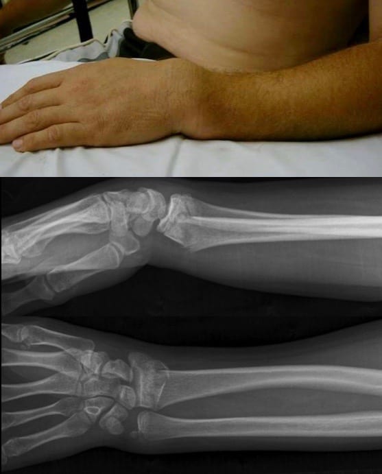
It’s painful, it presents acutely! And as Abraham Colles described, the wrist looks like a dinner fork, caused by dorsal swelling (caused by the distal fragment and local haematoma).
Examine the joint above and below
Document mechanism:
younger people will have a more marked mechanism of injury and the elderly a low energy mechanism of injury that suggests skeletal fragility.
Document neuro-vascular status, side of dominance and profession.
Don’t forget to remove any rings or watches from the affected side (and pop them on the unaffected side so they don’t get lost).
Analgesia at presentation:
I like to think about a ‘timeline of analgesia’. Oral analgesia is probably not going to help in the first hour, maybe oramorph works in 15 to 30 mins but oral analgesia should form a midpoint of the analgesic timeline.
Start with something that works immediately in the analgesia timeline: nasal diamorphine or fentanyl or inhaled agents such as Entonox or Penthrox.
If necessary give IV analgesia and of course apply a (elevation) sling.
Early use of inhaled analgesia: Penthrox or Entonox – allows for better images, rotated images affect the standard of fracture angulation measurement and xrays are sore to get right. #rcemblogs
– Rob Greig (@drrobgreig) July 2, 2020
You need to achieve a state of analgesia where the patient is comfortable and able to move their wrist onto the x-ray plates, which is the next stage of the radial fracture pathway.
X-ray: Get it right.
Inadequate analgesia gives you inadequate images which means inadequate management.
Get it right first time. Think efficiency of the X-ray department: you waste a finite resource if the patient has to return for better images because the initial analgesia wasn’t sufficient, and that slows the functioning of the ED.
You only need an AP and a lateral image.
FROM LITFL
X-ray: Description of fracture
Hopefully the X-ray confirms your clinical suspicions. When bones break during falls on outstretched hands invariably they give/fracture at one point.
Case courtesy of Dr Usman Bashir
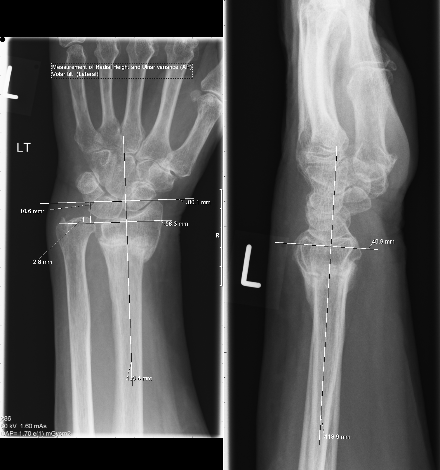
So how angulated is angulated? Well, draw a line across the C shape of the joint surface of the radius (on the lateral view) and run a perpendicular off from that line. Also on the lateral view, draw
a line along the shaft of the 1st metacarpal. The C-Radial line should point down the shaft of the 1st metacarpal (approximately). If the fracture is dorsally angulated you describe that angulation
relative to the metacarpal line.
So how short is short? Invariably the radial styloid distal point will be at the same level as the ulna styloid with a distal radial fracture. It should, in its typical anatomical position, be placed distal to it. For shortening to be described you look at the AP film. The radial articular surface adjacent to the ulna medial articular surface should be side by side. During distal fracture, the radial surface gets displaced (shortened) proximally. An even quicker method is just measure the overlap of the fracture segment on the remaining radial shaft.
There’s some really lovely diagrams describing the measurements here.
X-ray normal:
Classically if pain is out of proportion to the X-ray findings, i.e. the X-ray doesn’t show a distal radial fracture then you may have clinically missed something. Think scaphoid, forearm shaft, dislocation, other carpal fracture (like a cheeky dorsal triquetral fracture).
You cannot exclude a scaphoid fracture without scaphoid views and time. Be cautious, however scaphoid fractures are off the scope of this blog.
Now what?
So you have a distal radial fracture case. You have described its shortening and its dorsal angulation.
The patient is hopefully comfortable.
But do you plaster and send to orthopaedic clinic, manipulate and send to clinic or do you admit?
This is a tricky bit and needs experience. As a rule of thumb, if it’s short or angulated, it needs manipulating.
For every 10 degrees of dorsal angulation the likelihood of that fracture healing in its current position carries approximately a 10% chance of wrist disability. So if your patient is a concert pianist, get orthopaedics involved early.
If the fracture is intra-articular that needs discussion with orthopaedics as the ideal will be to try and close that joint surface gap. If the fracture is comminuted, it’s likely that a manipulation will fail to hold without K wiring.
But there are no absolutes, in my opinion. This is where the Rule of Sod appears: all the ones you think fail, succeed and the ones you think you’ll get a win on, fail! A similar thing happens when I aspirate spontaneous pneumothoraces.
Ok so you are going to reduce…
This is where even more opinions come out. This is my 20 year take on wrists.
Firstly manipulation is painful. You are effectively going to make their fracture worse before it becomes better.
Haematoma blocks vs Biers blocks vs Propofol/Ketamine vs Regional Blocks vs Penthrox.
I’m sure you all have an opinion and there is a paper indicating superiority of one of these versus another, with a recent one suggesting haematoma blocks are perhaps operator dependent. But it’s all operator dependent. I don’t care how you achieve analgesia, what I care about is that your patient doesn’t experience any more discomfort. The more comfortable the patient, the easier the reduction.
I’ve witnessed a haematoma block done so well that consultant literally jiggled the fragment after and patient was unbothered. Didn’t realise that was possible.
– Dr Prerna Chinoy (@preechinoy) July 2, 2020
I once amazed myself by doing one so good the lady held her arm aloft and waved it around with the hand and wrist just kinda flopping around.
I asked her to stop.– Andrew Jacklin (@Psynian) July 2, 2020
If you are skilled in all the above techniques great, if you are not then it’s an opportunity to learn new techniques or just focus on one technique.
I will say that haematoma blocks get a bad press, but they have a lead on the others and you’ll see why. The BOAST guidelines recommend not using haematoma blocks but they do list NICE guidelines, and BSSH guidelines as their evidence for saying so – neither of which say that! Entonox on its own has pretty much been outlawed by everyone.
I suggest you take a pragmatic approach to any of the above analgesic techniques:
Bear in mind department work load, patient type, patient choice of analgesic strategy, staff availability and availability of certain kit…
1. Haematoma Block: you can use PoCUS to guide, you can infiltrate into the haematoma, but I also infiltrate around the fracture periosteum. I have used chirocaine, chirocaine-lidocaine mix or just lidocaine. The key is to give the block time to work, many clinicians just rush in and are surprised that the patient experiences pain! Perform the block and then set everything else up whilst the block is “cooking”. Haematoma blocks also provide analgesia for after the manipulation, giving time for oral analgesia to catch up. Also if after the check X-ray a tweak is needed, it’s likely it’s still working (the “tweak window”). You can do this as a lone practitioner or have an untrained assistant.
Haematoma Block: How too
Firstly, check that a haematoma block is likely to work. If the fracture is old, there will be no haematoma to block – so a haematoma block won’t work. You’ll have to use some of the other options.
Without Ultrasound:
1. Feel for the fracture – if you look at the x-ray, then you know roughly where the break is. You can then palpate the fracture through the skin. It’s really useful to measure precisely if the wrist is so swollen you can’t feel.
2. Mark the fracture site with a pen.
3. Clean the area carefully
4. Draw up 5-20ml local anaesthetic (bear in mind your local anaesthetic toxicity doses). You’ll need lidocaine because it works quickly, but many people find bupivacaine useful to provide the volume. 5ml 2% lidocaine, 5ml 0.25% bupivacaine normally does the trick. The volume helps to distract (move apart) the fracture fragments.
5. Pierce the skin, and aim to get the tip of the needle in the haematoma from the dorsal surface. If you have about a 30 degree angle, it’s easier to get there. Aspirate as you go.
6. When you aspirate haematoma, inject your lidocaine.
How do you know it’s haematoma not blood? It’s dark, thick, red blood that sinks in your lignocaine syringe – and doesn’t really mix well.
With Ultrasound:
Ultrasound is useful to identify the haematoma, but also to check the post manipulation position before going to x-ray, incase further tweaks are needed.
There’s lots of resources – have a look at the this guide, and practice! We’ve not done a full literature review, but there is evidence suggesting these blocks work.
2. Biers block: you need prilocaine (although I have used lidocaine), time and equipment. You need a double cuff machine, the cuff is painful, you need a particular patient choice and resus monitoring. You also need at least one trained assistant.
Alas when you need the check X-ray, unless your X-ray team can perform the check X-rays in resus then you have to wait for the block to finish and lose the chance of tweaking your manipulation.
For full instructions, read the RCEM guideline, and practice under careful supervision.
BIERS BLOCK.
Better reductions. Fewer operations. Safe. Better for teaching. @Oceancurl66 recently had a look at our outcomes.
Just BETTER. #rcemblogs @CredeAndreas @casualtysrus @chrisconnolly83
– Tom Bircher (@tombircher) July 2, 2020
3. Propofol/Ketamine Procedural Sedation: in general, sounds very sexy. The flip side is you need more staff, have more risk and need more time. If you have the competency, another doctor with at least 1 nurse and you can give the patient time to recover, then great. Most departments on most days don’t have this luxury, and the patients often have lots of co-morbidities putting them into a higher ASA catagory. Plus you don’t have a tweak window post check X-ray.
4. Penthrox: 1 doctor and 1 nurse. I personally don’t have much experience with using Penthrox in wrist fracture reduction as a lone method. There are case reports. But you have a tweak window by reusing the Penthrox (as long as the max dose hasn’t been exceeded).
I find many elderly patients find using Penthrox for analgesia difficult. Attaching it to a face mask and coaching their breathing for at least fifteen minutes can help. People often don’t give it enough time to work
– Sandy Nelson (@sndynlsn) July 2, 2020
5. Hypnosis: OK, ok, I know you think I’m mad, and yes, this is an editor’s take over. But…there’s lots of case studies on using hypnosis to provide analgesia for joint manipulations, and also case studies on using hypnosis to promote fracture healing. Even just starting some 7-11 breathing can make a huge difference.
Fracture reduction.
So perfect analgesia has been achieved and the patient is relaxed (or unconscious if Propofol or Ketamine has been used)…
I don’t always have an assistant to help me reduce radial fractures and a brutish approach also disturbs me.
The best tool for reduction is your thumbs. Imagine you are holding the radius like a pen that you are snapping in two with your hypothenars on the ulna aspect of the wrist:
FIRST MAKE IT WORSE: you need to disentangle the fracture. The pieces of the fracture are like two cogs that are malaligned. So start by exaggerating the dorsal angulation. This is where you will feel crunching on your thumb pulps. Do not apply traction initially, it won’t reduce the fracture.
This is described in the EMJ, as the “Christmas technique” using hyper-flexion. We often use the opposite – hyperextension as described below.

Then you need to MAKE IT BETTER: here you apply some distal distraction and then volar angulation/palmar flex. This restores radial length and locks the angulation of the ‘fracture cogs’ together. This video (from about 2min in) demonstrates the bone movement – notice how the traction alone wouldn’t work.
Keep your thumb on the fracture ”lump” as you manipulate so that you can feel it disappear when you reduce it properly.
– Jim Crawfurd (@jim_crawfurd) July 2, 2020
Other techniques suggested for reducing Colles include finger traps or elevation. If you elevate a Colles by the fingers, the weight of the elbow will provide traction. If the patient doesn’t have much muscle bulk to resist this traction, it can work well – but, as the picture above shows, just traction might not be the best option.
Now plaster: plaster in ulna deviation and slight volar/palmar angulation.
Ensure the partial ‘backslab’ plaster envelops the distal radius dorsal, lateral and volar surfaces (Charnley Slab).
Just a dorsal ‘backslab’ is no good, the fracture reduction will slip.
Once the cast has set, document post-reduction neurovascular status. Put the arm in a sling and send for a check x-ray.
Ensure at discharge that the patient has adequate analgesia, orthopaedic follow-up and advice on possible issues with the plaster, excessive pain or paraesthesia.
Complications of a distal radius fracture
Median nerve injury either acutely or by delayed development of Carpal Tunnel Syndrome.
Vascular injury
Malunion
Manipulation failure, either acutely or late by fracture slip (often as the swelling subsides but also as the fracture site remodels)
Extensor Pollicis Longus (EPL) tendon rupture: painless, around the time the cast comes off in my experience.
Osteoarthritis
Chronic reduced wrist movement.
Grip strength reduction
We hope this has provided you with some tips and tricks for fracture manipulation. For those of you new to this – there’s nothing quite like finding an old hand to help you. For those of you who are very used to this – what other tips did we miss? Let us know.
Looking to improve your distal radius fracture reduction education? Check out this easy-to-make 3D printed simulator that facilitates procedural training by @wdixon1487 https://t.co/2I0UmyTc6Q #SBML #meded #3DPrinting @StanfordEMED #Simulation pic.twitter.com/jnB8APlyMg
— Perspectives (PME) (@pmeded) October 8, 2020

Foot Injuries
Foot injuries are common and because they affect mobility, they can have significant effects on a patient's life and so are important.

Elbow Injuries
Elbow injuries are common and acute elbow trauma accounts for 2-3% of all visits to the emergency department.

Zygomatic Complex and Nasal Injury
Anyone who has ever worked in an ED on a Friday night knows that facial injuries are a common presentation
20 Comments
Great summary. Our practice is to apply a full short-arm POP, with three point moulding on the second layer (normally over the reducers knee) with traction maintained throughout. The cast is then split when hardened. Anecdotally, seems not to slip as much and we don’t get folk reattending with blue fingers. Is this common practice elsewhere or do you all backslab?
Thank you for taking the time to write this very informative blog post. I would be grateful for clarification on the Christmas technique. On the link you have provided, it describes the first step as being ‘wrist hyperflexion’ to accentuate the dorsal angulation. My initial impression would have been to hyperextend the wrist to do this?
The Christmas technique article does indeed suggest hyper-flexion… however, as you say, we commonly use hyper-extension, as pictured. Either way a fracture fragment will be a pivot point. We might need to research which has better results!
Good review, focused
salient points nicely explained
very nice
Thank you, a good step by step approach
Great post with lots of valuable advice! Thank you
Wonderful take on this much needed topic!
straight forward and simple
Thanks. A practicle and very readable review.
Wow! great reduction technique
detailed and well documented. Like the drawings and markings.
Very readable and interesting
Great Summary. Have used haematoma block in practice but can appreciate that there is still some scepticism regarding its use from discussions with my consultants.
Great resource
As a new TACP I found this a really useful resource, easy to read and applicable to my practice
Great resource, thank. Have some experience in regional anaesthesia but definitely keen to try out a haematoma block
Great review
Reduced a Colles’ yesterday using only Penthrox – which before reading this article, I thought was a regular thing!