
Authors: Darren Davies, Remy Toko / Editors: Maya K Naravi, Helen Yasmin Sultan / Reviewer: Jon Bailey / Codes: NeuC10, NeuC14, NeuP8, NeuP9, SLO1, SLO5 / Published: 25/01/2021

Bell’s palsy is an idiopathic unilateral lower motor neurone facial nerve paralysis of acute onset [1] which includes:
- Inability to wrinkle the forehead on the affected side
- Inability to close the affected eye
- Flattening of the nasolabial fold on the affected side
The incidence of Bell’s palsy is:
- 20-25/100 000 per year in adults [2,3]
- 13-30/100 000 per year in all age groups [4]
- 1.6/100 000 per year in children [4]
There are no geographic, gender or race differences [2].
The incidence in children is lower than in adults and needs careful assessment to look for potentially serious causes [3].
Learning bite
Lower motor neurone paralysis of the facial nerve affects the entire face. Upper motor neurone weakness only affects the lower half of the face [1].
Anatomy
| Muscles supplied | Sensory nerve distribution | Parasympathetic nervous system functions |
| Facial expression | Taste – anterior two thirds of the tongue | Submandibular gland |
| Scalp | External auditory meatus | Sublingual gland |
| External ear | Soft palate | Lacrimal gland |
| Buccinator | Adjacent pharynx | Nasal |
| Platysma | Palatine glands | |
| Stapedius | ||
| Stylohyoid | ||
| Posterior belly of digastric |
The facial nerve (cranial nerve VII) supplies motor branches to the muscles of facial expression and supplies some sensory and parasympathetic fibres (see Table 1).

Bell’s Palsy and Other Causes of Facial Nerve Paralysis
Diagnosis of Bell’s palsy is reached only once other causes are excluded. In children, Bell’s palsy is a much less common cause of lower neurone facial nerve paralysis than is the case in adults.
Other causes of facial nerve paralysis may include:
Infection
|
Infections and facial nerve paralysis |
|
|---|---|
| Viral – Ramsay Hunt syndrome [6] |
|
| Lyme disease |
|
| Bacterial |
|
Trauma [5]
VII cranial nerve is the most commonly injured nerve in head trauma:
- Typically, temporal fracture with nerve transection
- Basal skull fracture [6]
Systemic diseases [5]
Systemic diseases which may cause facial nerve paralysis include:
- Sarcoidosis [5]
- HIV [5,6]
- Polio [6]
- Tuberculosis [6]
- Multiple sclerosis [6]
- Guillain–Barré syndrome [6]
- Diabetes [6]
Neoplasm [6]
Typically progressive course over 3 weeks, but sudden onset does not rule out:
- Parotid gland tumours [5,6]
- Pontine tumours [6]
- Acoustic neuroma [6]
- Leukaemia
Developmental hypoplasia/aplasia [7]
Learning bite
Bell’s palsy is only one of many potential causes of facial nerve paralysis [2,8].

Symptoms and Associated Features
A detailed history of symptoms and associated features should be reported.
Symptom onset includes:
- Date of onset [1]
- Rapidity of progression [1]
- History of preceding viral illness or trauma [1]
- Rash [3]
- Other evidence of infection
Associated features may include:
- Ear pain [3]
- Sensory changes on affected side of face
- Reduced tears [3]
- Overflow of tears
- Hyperacusis [3]
- Altered taste sensation [3]

Progression of paralysis beyond 3 weeks suggests a possible neoplastic cause and this is one of the reasons that follow-up is recommended for children presenting with facial nerve paralysis [2,3,8,9].
The MRI is of a 7-year-old boy with astrocytoma causing facial weakness. Click on the MRI scan to enlarge.
Neoplastic origin in children is suggested by [8,9]:
- Gradual progression of paralysis beyond 3 weeks [2,3]
- No return of function after 6/12
- Ipsilateral recurrence [2,3]
- Facial hyperkinesia (particularly hemifacial spasm)
- Associated cranial neuropathies and other neurological signs [2,3]
- Pain
- Single branch involvement
Neoplastic Origins
| Facial nerve paralysis | |
| Central | Upper motor neurone implies a cause other than Bell’s palsy |
| Peripheral | Lower motor neurone [8] |
The assessment of the child with facial nerve paralysis requires a full and thorough head to toe examination to ensure important clues to possible causes are not missed [10].
It is important to distinguish between the following types of facial nerve paralysis (Table 1):
- Central facial nerve paralysis
- Peripheral facial nerve paralysis
Neurological examination should include:
- Cranial nerves and peripheral nervous system
- Testing of VIIn should reveal a lower motor neurone (LMN) pattern with inability to wrinkle the forehead. Preservation suggests a central cause
The assessment of the child with facial nerve paralysis requires a full and thorough head to toe examination to ensure important clues to possible causes are not missed [10].
Examination of the ears includes:
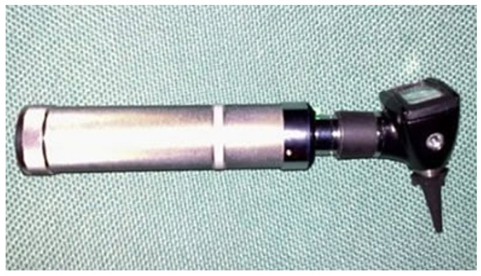
Examination of the ears with an otoscope and tuning fork testing [3] is critical, otherwise the rash of Ramsay Hunt syndrome may be missed.
Detailed testing of hearing using the Rinne and Weber tests is useful to identify the presence of other cranial nerve abnormalities which suggest the necessity for further investigation at the outset.
Rinne test

- Rinne test is conducted by tapping a 512 Hz tuning fork and holding it adjacent to the ear (air conduction), followed by placing the base of the tuning fork to the mastoid process (bone conduction)
- Normally, air conduction>bone conduction, i.e. Rinne positive [11]
- In conductive deafness, bone conducts>air (reversed), and Rinne test is positive with neural deafness [11]
Weber test
- Weber test is conducted by tapping a 512 Hz tuning fork and holding the base against the vertex in midline
- In neural deafness, the tone will be heard better in the intact ear [11]
- In conductive deafness, it is heard better in the affected ear [11]
Examination of the face

Facial examination should include facial expression, inspection of the oropharynx for asymmetry [3], assessment of eye closing and inspection of the eye itself for inflammation, tearing, pain and visual impairment. Where the eye cannot be closed, fluorescein instillation and examination under cobalt blue light should be performed to exclude corneal abrasion and ulceration [1]
Rashes

Examine the skin for rashes. Specific relevant rashes include Erythema migrans, which may suggest Lyme disease (see photo), and Herpes zoster, which may suggest Ramsay-Hunt syndrome
Lymphadenopathy
Lymphadenopathy may be present with lymphoma or neoplasm and is an indication for specialist referral.
Blood pressure
Hypertension, when identified with facial nerve paralysis, requires specialist management and further investigation.
Joint examination
Examine the musculoskeletal system for pain and swelling, particularly in the major joints. Joint involvement may be seen with Lyme disease
A history typical of Bell’s palsy with normal findings on examination does not warrant investigation immediately.
Most children presenting with uncomplicated facial nerve paralysis need no investigation.
However, there are exceptions. Some ENT and Haematological specialists recommend performing a full blood count with film in all cases at the outset to identify serious causes such as leukamia early [2,12].
Children with a history of travel to or living in an area where Lyme’s disease is endemic and a history of exposure to a tick prone environment such as field or forest, should have Lyme serology performed.
Lyme disease distribution can be found here.
Suggestive history, central or UMN CNVII palsy, other focal neurology peripherally, or other cranial nerve abnormality on examination should all prompt imaging. MRI is preferable for investigating a possible underlying neoplasm [1]; CT is preferable if an acute traumatic cause is suspected.
Formal follow-up is essential, irrespective of investigations performed. Nerve conduction studies, electromyography and electroneurography, and acoustic reflex determination (ipsilateral and contralateral) are not investigations required in the emergency department, however, these may have a role during follow-up [2].

Features atypical of Bell’s palsy require referral18 for exclusion of an alternative diagnosis and include:
- Insidious and painful onset. Gradual progression is more likely to be associated with a neoplastic or infectious cause of facial palsy.
- A progressive and prolonged (more than 3 months) duration of symptoms with frequent relapses (indicative of a neoplastic process).
- Predisposing factors for facial palsy, for example, previous stroke, brain tumour, parotid tumour, skin cancers of the head and face, or facial trauma.
- Systemic illness or fever.
- Vestibular or hearing abnormalities (other than hyperacusis), otorrhoea, diplopia or dysphagia.
- Sparing of forehead movement (which may indicate an upper motor neurone lesion such as stroke) and bilateral signs (may be indicative of Lyme disease or sarcoidosis). Lower motor neurone lesions (such as Bell’s palsy) do not spare the upper face.
- A recurrent episode.
- Paralysis of individual branches of the facial nerve or other cranial nerve involvement.
- Parotid gland masses, vesicular skin rashes, and lesions suggestive of skin cancer.
Refer to a facial nerve specialist (for example, a neurologist or ear, nose, and throat specialist) if there is doubt about the diagnosis or a person with Bell’s palsy has:
- No improvement after 3 weeks of treatment.
Management of Bell’s Palsy
There is very little evidence specifically from paediatric populations on the management of Bell’s palsy, despite a growing adult evidence base.
Corticosteroids
In the management of adult Bell’s Palsy, following a Cochrane review in 2015 there is a clear recommendation to use corticosteroids if the patient presents within 72 hours of the onset of symptoms. The dosage regimen, however, remains unclear.
In the paediatric population, there remains an absence of evidence. Neither a 2005 BestBET review nor a 2012 systematic review were able to identify any randomised controlled trials addressing this question. A protocol for BellPIC, a multicentre, placebo-controlled randomized trial, aims to answer this question by 2020.
Follow-up Strategies
If no other cause of acute facial paralysis is identified, in the absence of risk factors for underlying malignancy or other causes, the patient may be discharged from the ED with a suitable follow-up plan:
GP

- Ideally the child should be seen within 3 weeks by their GP or a doctor in the ED for review
- If, at this stage, there are any signs of progression or any other worrying features have become apparent, then referral to a specialist should be made and arrangements made for further testing (electro/neurophysiology testing)
Follow-up
- If there are signs of recovery by 1 month, routine follow-up at 3, 6 and 9 months is recommended [1]
- Monitoring should be for a minimum of 1 year (risk of missing long term sequelae if not done) [3]
Prognosis
- Examination to determine if paresis (incomplete paralysis) or complete paralysis is present is the most important prognostic test at the outset
- Paresis at onset can be followed up clinically, because prognosis is invariably good
- Further testing is recommended in cases of complete paralysis or if no signs of recovery within 3 weeks of disease onset [1]
- In patients who recover without treatment, major improvement occurs within 3 weeks in most cases [3]
Natural history of Bell’s palsy in children states that [17]:
“True Bell’s palsy is thought to be more benign, with a tendency toward complete resolution in many cases within 2 months of onset of facial paralysis and by 6 months in most cases.”
Discharge advice needs to contain information on worrying features so that parents/patients themselves can return early if such features develop [10].
Upon discharge, the following should be explained:
- Administer analgesia for pain as needed (paracetamol)
- Child may experience sensitivity to sounds during acute stages
- Child may initially worsen before improving
- Most children recover fully by 6 weeks, although may take up to 1 year
- 10% will get some continuing weakness or the nerve may grow back to the wrong area
- Medical advice should be sought if any concerning symptoms occur
Concerning symptoms
- Red/painful eye
- Progression of weakness after 48 hours
- Different or new symptoms such as:
- Headache
- Vomiting
- Temperature
- Disturbed vision
- Weakness
- Abnormal sensation in another area of the body or head or neck
- No improvement in facial paralysis after 1 month
- Singhi P, Jain V. Bell’s Palsy in children, Seminars in Pediatric Neurology 2003;10(4):1071-9091.
- Pancioli A. Part Three Medicine and Surgery, Section VII Neurology. In: Rosen, ed. 99 Brain and Cranial Nerve Disorders: Facial Nerve Paralysis. 5th Ed Vol 2. Oxford: Oxford University Press, 2006:1488-1489.
- El-Haurani AS, Eng CY, Ahmed SK et al. General Practitioners referral pattern for children with acute facial paralysis, Journal of Laryngology and otology 2005;199(7):0022-2151.
- Wyatt JP, Illingworth RN, Grahm CA et al. Chapter 12: Ear, Nose and Throat – Facial nerve palsy In: Oxford Handbook of Emergency Medicine 3rd Ed. Oxford: Oxford University Press, 2006:552.
- Hope RA, Longmore JM, McManus SK et al. Chapter 10: Neurology, Cranial Nerve Lesions Bell’s Palsy. In: Oxford Handbook of Clinical Medicine. 4th Ed. Oxford: Oxford University Press, 1998:460-461.
- Polin R, Ditmar M. Pediatric Secrets. 2nd Ed. Philadelphia: Mosby, 1997.
- Grundfast KM, Guarisco JL, Thomson JR et al. Diverse etiologies of facial paralysis in children. International Journal of Pediatric Otorhinolaryngology 1990;19(3):0165-5876.
- Lunan R, Nagrajan L. Bell’s palsy: a guideline proposal following a review of practice. Journal of Paediatrics & Child Health. 2008;44(4) (219-20):1440-1754.
- Thomas J, Monaghan T. Chapter 10: The Nervous System Cranial nerve VIII: vestibulocochlear. In: Oxford Handbook of Clinical Examination and Practical Skills. 4th Ed. Oxford: Oxford University Press, 1997:314.
- Bhattacharyya AK, Ghosh S. Paediatric facial paralysis. Current opinion in Evaluation and Management. Indian Journal of Otolaryngology 1999;51:21-27.
- Ciorba A, Corazzi V, Conz V, Bianchini C, Aimoni C. Facial nerve paralysis in children. World Journal of Clinical Cases. 2015;3(12):973-979. doi:10.12998/wjcc.v3.i12.973.
- Ashtekar CS, Joishy M, Joshi R Do we need to give steroids in children with Bell’s palsy? Emergency Medicine Journal 2005;22:505-507.
- Pitaro J, Waissbluth S, Daniel SJ. Do children with Bell’s palsy benefit from steroid treatment? A systematic review. Int J Pediatr Otorhinolaryngol. 2012;76:921–926.
- Babl FE, Mackay MT, Borland ML, et al. Bell’s Palsy in Children (BellPIC): protocol for a multicentre, placebo-controlled randomized trial. BMC Pediatrics. 2017;17:53. doi:10.1186/s12887-016-0702-y.
- Madhok VB, Gagyor I, Daly F, Somasundara D, Sullivan M, Gammie F, Sullivan F. Corticosteroids for Bell’s palsy (idiopathic facial paralysis). Cochrane Database of Systematic Reviews 2016, Issue 7. Art. No.: CD001942. DOI: 10.1002/14651858.CD001942.pub5
- Gagyor I, Madhok VB, Daly F, Somasundara D, Sullivan M, Gammie F, Sullivan F. Antiviral treatment for Bell’s palsy (idiopathic facial paralysis). Cochrane Database of Systematic Reviews 2015, Issue 11. Art. No.: CD001869. DOI: 10.1002/14651858.CD001869.pub8
- Teixeira LJ, Valbuza JS, Prado GF. Physical therapy for Bell’s palsy (idiopathic facial paralysis). Cochrane Database of Systematic Reviews 2011, Issue 12. Art. No.: CD006283. DOI: 10.1002/14651858.CD006283.pub3
- National Institute for Health and Care Excellence (NICE), Bell’s palsy: NICE CKS. Last revised: May 2019

Acute Facial Palsy
This session looks at the assessment and management of acute facial palsy.

The Droopy Dribbler: Paediatric Presentations of Bell’s Palsy
Paediatric cases of Bell's Palsy are relatively uncommon (6.1/100000 in the age range 1-15 (1)); understandably, witnessing a rapidly developing facial asymmetry in a child will cause worried parents/guardians to rush to see a doctor

Common Entrapment Syndromes
Nerve entrapment syndromes are a group of conditions in which peripheral nerves are damaged, through compression or repeated trauma.
9 Comments
Excellent overview of management of Facial Nerve Palsy
excellent
great summation of bells palsy
Excellent
very useful
Excellent case.
very useful
Very useful overview. Thanks.
very useful and practical review.