
Author: Marie-Clare Harris / Editor: Marie-Clare Harris / Reviewer: Mehdi Teeli / Codes: C3AP1b, CAP1, CMP3, HAP1, HMP3 / Published: 15/09/2020
This session will give you a plan for the initial assessment and management of abdominal trauma. It should enable you to think ahead and try to predict what underlying intra-abdominal injuries a patient may have sustained.
It is a good idea to be familiar with the trauma services available in your hospital. The imaging services, radiological interventions and surgical capabilities will vary between a regional trauma centre and a district general hospital. If you are working in a department where the most appropriate services are not available on site, it is crucial to start thinking early about how best to access those required.
Before commencing this session you should have:
- A good knowledge of the anatomy of the abdomen and the organ systems contained within
- An understanding of the physiology of hypovolaemia and shock, and the body’s response to pain and injury
Context
The assessment of abdominal trauma is notoriously difficult.
Serious intra-abdominal injuries may initially appear to be innocuous and localisation of symptoms is variable. A high index of suspicion is constantly required to recognise occult injury. The presence of associated injuries, such as head injury with altered conscious level, spinal injury with neurological sequelae or adjacent chest or pelvic injuries, further complicates clinical evaluation.
To aid initial diagnosis, investigation and management, abdominal trauma can be broadly grouped into penetrating or blunt, depending on the mechanism of injury.
Learning Bite
Unrecognised abdominal injury remains a significant cause of death. Death may be prevented if intervention occurs early.
Penetrating Trauma
In penetrating trauma, the injuries sustained are caused by direct injury as the penetrating item passes through the organs and tissues in its path.
In the UK, this is most commonly knife-related injury. The site of entry, direction of the injury and the size and nature of the penetrating item will all provide information regarding the underlying injuries. The visible wound may be small and insignificant in appearance.
Stab wounds and low-energy gunshot wounds cause tissue damage by lacerating and tearing. High-energy gunshot wounds transfer more kinetic energy, causing increased damage surrounding the track of the missile due to temporary cavitation.¹

Learning Bite
It is impossible to tell from the appearance of a surface wound, the extent of underlying damage.
Wound Assessment
Caution is required in assessment of penetrating abdominal wounds. Patients with penetrating injury, even with normal vital signs, should be assessed by a senior surgeon. Unless the injury is trivial, all patients should be admitted to hospital for observation even if no further investigation is initially required.
Do not remove any object remaining in the wound, such as a knife, as this may be tamponading underlying vessels.
Learning Bite
In a physiologically normal patient without signs of peritonism, wound exploration should only be undertaken by an experienced surgeon, ideally in theatre.
Common Sites of Injury
Approximately 50% of penetrating abdominal injuries damage intraperitoneal structures. The peritoneal cavity is not breached in 25% of cases.
Stab wounds traverse adjacent abdominal structures and most commonly involve the liver (40%), small bowel (30%), diaphragm (20%), and colon (15%).
Blunt Trauma
Blunt abdominal trauma commonly results from either a compression force or a deceleration injury. It has greater mortality than penetrating as there is often injury to multiple organ systems.
There are a number of classical injury patterns, which occur following common mechanisms of injury such as rapid deceleration while wearing a lap-only seat belt, or after direct impact with a car steering wheel. Recognition and understanding of these patterns will aid subsequent assessment.
The spleen and liver are the most commonly injured intraperitoneal organs in blunt trauma.
Injury to intraperitoneal structures should be considered when evaluating any injury which occurs between the nipple line and the pelvis.
Abdominal Contents
Detailed anatomical knowledge of the abdomen is central in enabling the clinician to systematically assess patients with abdominal trauma and predict likely sites of injury.
The abdominal contents can be divided into:
- The intra-abdominal structures
- Retroperitoneal structures
- The pelvic structures
Learning Bite
Detailed anatomical knowledge will enable an accurate appreciation of likely organ damage.

Fig 1: The layout of the intra-abdominal contents

Fig 2: The retroperitoneum and the structures contained within it

Fig 3: The great vessels of the abdomen and pelvis
Abdominal Anatomy
Injury to any region of the abdomen can of course create injury to any of the organs contained therein. However, commonly injury to a specific area will result in injury to those organs contained within that locality.

Fig. 4
| Region | Organs potentially injured |
| Lower chest | Liver, spleen, diaphragm, stomach |
| Anterior abdomen | Liver, spleen, colon, bladder, stomach, pancreas, transverse colon, ileum, jejunum |
| Flank | Kidneys, ureters, ascending and descending colon |
| Posterior abdomen | Great vessels, duodenum, pancreas, spinal cord |

Image 1: Organs of the lower chest (mid to lower thoracic cage) 15% of stab wounds to this area are associated with significant visceral damage

Image 2: Organs of the anterior abdomen (between anterior axillary lines) 60% of injuries to this region penetrate the peritoneum and, of these, 4050% cause visceral damage

Image 3: Organs of the flank (between anterior and posterior axillary lines)
Injury Signs and Symptoms
Symptoms and signs are normally related to blood loss from injury to these very vascular organs. Signs of shock may be present but will be a late sign in a young, fit patient.
Injury to the liver or spleen are common injuries following blunt trauma and can be identified on a trauma CT scan.

Image 1: Signs and symptoms of a patient in hypovolaemic shock
Liver
Liver injuries account for 15-20% of intra-abdominal organ injuries but up to 50% of mortality, and 45% have associated splenic injury.
Conservative management is appropriate in 80% of cases, with surgical intervention reserved for ongoing and uncontrolled bleeding.
If hepatic or splenic injuries are detected on CT, the source of any ongoing bleeding can be detected through angiography. Through interventional radiology it can be possible to embolise the bleeding vessel and remove the need for surgical intervention.

Image 2: CT scan showing laceration to the liver
Spleen
Splenic injury is graded according to CT findings and treatment is guided by grade:
|
Grade 1
|
Minor subcapsular tear or haematoma
|
|
Grade 2
|
Parenchymal injury not extending to the hilum
|
|
Grade 3
|
Major parenchymal injury involving vessels and hilum
|
|
Grade 4
|
Shattered spleen
|
The spleen contains approximately one unit of blood at any time.
Grade 1 or 2 injuries can usually be managed conservatively.

Image 3: CT scan showing ruptured spleen
Pancreas
- Injury to the pancreas may cause pancreatitis, which may develop over days.
- Blunt pancreatic injury may not be immediately recognised. It is relatively uncommon, occurring in around 10% of blunt abdominal injuries but it is rarely an isolated injury due to the position of the pancreas.
- Amylase elevation will often not occur until 3-4 hours after injnury, if at all, and lipase is no more specific for pancreatic trauma.
Other Organs
The hollow organs, vascular structures and genito-urinary (GU) tract can also be injured and will each display their own characteristic responses to injury.

Image: Onset of peritonitis
Hollow viscus
- Peritoneal contamination with bowel contents will produce peritonism.
- There may be accompanying blood loss but the degree of hypovolaemia is generally less significant than in solid organ injury.
- Damage to the retroperitoneal portion of the bowel will not produce classical signs of peritonism as the leak will be contained.
Vascular structures
- Catastrophic blood loss may occur with injury to any of the large vessels in the abdomen.
- Aortic injury is usually fatal, but may be tamponaded if it occurs retroperitoneally.
- Injury to the inferior vena cava is likely to be associated with more insidious blood loss unless there is a large tear.
GU tract
- Bruising, haematuria or meatal blood are often the only signs of a GU injury.
- Injury to the intraperitoneal portion of the bladder may result in chemical peritonitis.
As with all trauma management, the priority is to identify immediately life-threatening injuries. Early airway protection, ventilatory support and circulatory resuscitation are paramount. An accurate history, if possible, will guide subsequent management. You may be reliant on the testimony of eyewitnesses or the ambulance service.
Clinical examination can be unreliable and falsely reassuring in the multiply injured patient. A significant mechanism and injury to adjacent body cavities should be taken seriously and prompt a thourough search for abdominal trauma.

Analgesia
Patients with abdominal trauma may require significant resuscitation and analgesia prior to formal abdominal assessment. In all cases where there is suspicion of abdominal trauma and blood loss, an urgent cross-match sample must be sent.
In haemorrhagic shock, the priority is resuscitation to restore adequate tissue perfusion and oxygen delivery. Adequate intravenous analgesia should be administered promptly and not delayed whilst investigations are obtained.
Learning Bite
A patient should always receive adequate intravenous opioid analgesia titrated to clinical response.

Image: Fluid replacement therapy and analgesia for the patient in hypovolaemic shock
Management of Haemorrhagic Shock
Normal saline is the recommended initial intravenous resuscitation fluid. High flow oxygen and blood products, if appropriate, will maximise the oxygen-carrying potential of the intravascular fluid.
Management of haemorrhagic shock involves restoration and maintenance of circulating volume and tissue perfusion until bleeding is controlled. A patient who is GCS 15 and physiologically normal can be viewed as being adequately resuscitated, although frequent reassessment of this state is required. A patient who is initially haemodynamically normal and has no sign of significant abdominal injury can be treated in an observant fashion.
The table below shows the grades of hypovolaemic shock.
| Grade I | Grade II | Grade III | Grade IV | |
| Blood loss | <750ml | 750-1500ml | 1500-2000ml | >2000ml |
| Pulse | normal | tachy | tachy | weak |
| Blood pressure | normal | raised diastolic | lowered systolic | very low |
| Mental state | normal | agitated | confused | obtunded |
| Urine output | 30ml/h | 20-30ml/h | 5-15ml/h | <5 ml/h |
| Respiratory rate | normal | normal | tachypnoeic | tachypnoeic |
Fluid Resuscitation
Aggressive fluid resuscitation without adequate haemostasis has been demonstrated to dilute clotting factors and potentially dislodge clots. Maintaining a normal blood pressure may worsen bleeding. If there is ongoing and brisk bleeding, it may be impossible to adequately resuscitate a patient until the tap is switched off.
Examples of this situation would be a significant liver and spleen injury, or injury to one of the large intra-abdominal vessels. Continued fluid administration in this setting will eventually serve to haemodilute and impair coagulation. Immediate CT should be carried out and transfer from there to the angiographic suite if available or operating theatre depending on findings.
Learning Bite
Continuing abnormal physiology despite aggressive fluid resuscitation mandates early surgical intervention to turn off the tap.
Clinical examination of the abdomen should always be undertaken as part of the primary survey. A conscious patient may complain of abdominal pain but in other cases, a patient with a distracting injury may less aware of their abdominal injury.examination findings may be minimal even in significant injury and an index of suspiscion must be maintained.
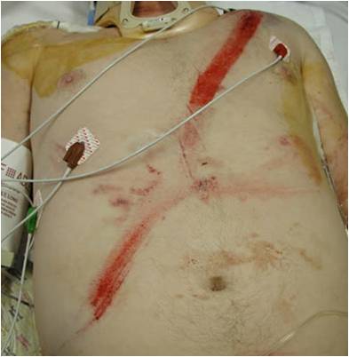
Inspection of the injured abdomen may show evidence of bruising, abrasions or penetrating trauma. Abdominal distension is suggestive of intra-abdominal haemorrhage and in association with signs of hypovolaemia must be dealt with promptly.
Palpation of the abdomen should be carried out and patient response documented. It is of particular importance to palpate the renal angles and examine the external genitalia.
Tenderness or guarding in any region must be noted and further investigated, but a multiply injured patient may not report much tenderness to palpation even with underlying injury.
A rigid abdomen is due to leak of bowel contents into the peritoneum from a hollow viscus injury.
Blood is by itself not a peritoneal irritant and, in a patient who has undergone a significant injury, serial examinations may demonstrate a gradually distending abdomen with little initial discomfort.
Rectal examination is useful if it demonstrates bleeding or a high-riding prostate suggestive of a pelvic injury and involvement of the genitourinary tract. Anal tone and sensation should be tested however,you should be aware though that in a head or spinally injured patient these will not be appreciated and cannot be used to exclude a pelvic or neurological injury.
Top-to-toe examination including a log-roll is imperative to ensure there are no other injuries.
Whilst it is vital to fully expose the patient to adequately visualise all areas, attention should be paid to temperature control. Hypothermia in trauma patients is associated with significant mortality.
Learning Bite
Remember that an unconscious or spinal-injured patient will not be able to feel or localise abdominal tenderness.
A definitive patient management plan should be formulated as early as possible, including early consultation by relevant speciality teams. This may include surgeons, radiologists, anaesthetists, theatre staff, critical care and transfer teams if definitive care is not available in your hospital.
In a trauma centre, early tramua CT should be carried out, ideally once the patient has been haemodynamically stabilised through fluid resuscitation. If a patient remains unstable despite resuscitation then senior members of the trauma team (emergency, surgical and anaesthetics) should make a team decision weighing up the merits of CT versus immediate theatre.
In a DGH, contacting radiology to arrange imaging and thinking about arranging transfer to a trauma centre should be done as early as possible.
Trauma
Patients with suspected abdominal injuries from trauma can be roughly categorised into three groups:
- Those requiring immediate surgery due to catastrophic haemorrhage unresponsive to resuscitation
- Those requiring urgent investigation using appropriate diagnostic tests to determine intra-abdominal injury and decide the most appropriate management
- Those with no abdominal injury on clinical examination and where there is a low index of suspicion who require observation and sequnetial re-examination.
Patients who are physiologically normal and who have no indication for immediate imaging or intervention should be discussed early with the surgical team. A decision can then be made regarding the appropriate location for further observation.
Fluid resuscitation
Choice of fluid: isotonic crystalloid solution, up to 1000ml in adults, 20ml/kg in children.
| table 3-2 responses to initial fluid resuscitation∗ | |||
| RAPID RESPONSE | TRANSIENT RESPONSE | MINIMAL OR NO RESPONSE | |
| Vital signs | Return to normal | Transient improvement, recurrence of decreased blood pressure and increased heart rate | Remain abnormal |
| Estimated blood loss | Minimal (<15 % ) | Moderate and ongoing (15%–40%) | Severe (>40%) |
| Need for blood | Low | Moderate to high | Immediate |
| Blood preparation | Type and crossmatch | Type-specific | Emergency blood release |
| Need for operative intervention | Possibly | Likely | Highly likely |
| Early presence of surgeon | Yes | Yes | Yes |
| ∗Isotonic crystalloid solution, up to 1000 mL in adults; 20 mL/kg in children | |||
Adapted from atls manual 2010
| comparison of dpl, fast, and ct in abdominal trauma | |||
| DPL | FAST | CT SCAN | |
| Advantages |
|
|
|
| Disadvantages |
|
|
|
| Indications |
|
|
|
Adapted from atls manual 2010
Key Learning Points
- Abdominal trauma is an important but often preventable cause of mortality.
- Be alert to clues from the paramedics. Early recognition and initiation of appropriate treatment is crucial.
- Consider that all trauma patients have intra-abdominal injury until proven otherwise.
- Adequate resuscitation and frequent reassessment are vital, but definitive surgical intervention may be required. Involve the surgical team early.
- Start thinking ahead. If your patient is likely to need an operation, alert the appropriate personnel as soon as possible.
Safety Pearls and Pitfalls
- The presence of a physiologically normal patient does not exclude significant intra-abdominal injury
- Intoxication, head injury or distracting injuries may make clinical assessment of the abdomen unreliable
- An unconscious, anaesthetised or spinally-injured patient will not display signs of guarding
- Flank injuries may produce retroperitoneal injury to the kidneys or bowel without any initial symptoms. Damage to this area should prompt a search for such injuries
- Intraperitoneal blood will not produce signs of peritonitis
- A high-risk mechanism of injury dictates that a patient should be admitted and re-examined over several hours
- Always expose and inspect the entire patient. Just because you have found one stab wound doesn’t mean there’s not another one
- Beware of penetrating buttock injuries
- If there is an implement still in situ, leave it alone.
- AMERICAN COLLEGE OF SURGEONS. (2004) Advanced trauma life support (ATLS) for doctors, 10th ed. Chicago: American College of Surgeons.
- ARGALL, J. (2002) Factor VIIa for intractable blood loss in trauma [online] BestBETs Available from: www.bestbets.org/cgi-bin/bets.pl?record=00371 [Accessed 24th March 2009].
- BROOKS, A.J. and ROWLANDS, B. J. (1999) Blunt abdominal injuries. British Medical Bulletin, 55, p. 844-855.
- DUTTON, R.P. et al. (2002) Hypotensive resuscitation during active haemorrhage: Impact on in-hospital mortality. Journal of Trauma-Injury Infection & Critical Care, 52(6), p. 1141-1146.
- GARNER, J. (2005) Blunt and penetrating trauma to the abdomen. Surgery 23(6), p. 223-228.
- GERMANOS, S. et al. (2008) Damage control surgery in the abdomen: an approach for the management of severe injured patients. International Journal of Surgery, 6(3), p. 246-252.
- HAYES, C.W. et al. (1991) Seat belt injuries: radiologic findings and clinical correlation. Radiographics, 11(1), p. 23-36.
- LAWSON, R. B. and GOOSEN, J. (2009) Abdominal Stab Wound Exploration [online], eMedicine. Available from: http://emedicine.medscape.com/article/82869-overview [Accessed 24th March 2009].
- MARX, J.A. et al. (2005) Rosens Emergency Medicine: Concepts and Clinical Practice, 6th ed. Philadelphia: Elsevier Health Sciences
- NARANG, S. (2005) Is conservative management of stab wounds better than wound closure? [online] BestBETs Available from: www.bestbets.org/bets/bet.php?id=00581 [Accessed 24th March 2009].
- PINHEIRO, L. (2005) Infrahepatic Inferior Vena Cava Injury [online] Trauma.Org. Available from http://www.trauma.org/index.php/main/case/17 [Accessed 24th March 2009].
- ROYAL COLLEGE OF SURGEONS OF ENGLAND and THE BRITISH ORTHOPAEDIC ASSOCIATION (2000) Better Care for the Severely Injured, Gosport: RCSENG Professional Standards and Regulation.
- SALOMONE, J. A. and SALOMONE, J. P. (2007) Abdominal Trauma, Blunt [online], eMedicine. Available from: http://emedicine.medscape.com/article/821995-overview [Accessed 24th March 2009].
- SHUMAN, W. P. and HOLTZMAN, S. R. (2005); ACR Appropriateness Criteria blunt abdominal trauma. [online] National Guideline Clearinghouse. Available from: www.guideline.gov/summary/summary.aspx?ss=15&doc_id=8270&nbr=4602 [Accessed 24th March 2009].
- STANTON-MAXEY, K. J. and BJERKE, H. S. (2007) Abdominal Trauma, Penetrating [online], eMedicine. Available from: http://emedicine.medscape.com/article/433554-overview [Accessed 24th March 2009].
- TINTINALLI, J.E., KELEN, G.D. and STAPCZYNSKI, J.S. (2003) Emergency Medicine: A Comprehensive Study Guide, 6th ed. New York: McGraw-Hill Professional.
- VIDYASANKAR, V., PHERWANI, A. D. and HANNON, R. (2003) Injuries to the abdomen and pelvis. Surgery, 21(8), p. 185-189.

Thoracic Trauma
For each life-threatening thoracic injury this session includes: a definition and context, Clinical assessment , treatment and Key learning points.

Abdominal Pain without Shock
This article covers the generic assessment and management of the pain with abdominal pain without shock.

Ultrasound in Emergency Medicine - Level 1 Instruction
This document covers FAST, Assessment of the Abdominal Aorta and IVC, Vascular Access and Echocardiography in Life Support.
38 Comments
Excellent topic
thank you ,nice topic .
Good topic
Complete overview of Management of Abdominal trauma.
Excellent module
Very nice topic
interesting read
very informative topic
Very useful and informative about abdominal trauma management in the emergency department.
excellent module on abdominal trauma.
very well taught thanks
Succinct article covering key points logically
Good overview of abdominal trauma.
Excellent topic
good module
good abdominal trauma overview
Very nice topic
It was well written. Points to major areas never to miss.
Excellent module- thanks
Good overview of abdominal trauma
Brilliant stuff
well presented and clinically relevant, good points about pitfalls and importance of trauma team input at early stage to avoid unnecessary delay of manamgement
Thorough and informative
Covered almost every aspect of traumatic abdominal injury.
nice , to the point
Revision of ATLS protocols in abdominal trauma
very valuable information
Succint yet comprehensive
Excellent overview of Abdominal trauma.
very good review of abdominal trauma
good revision
nice
A Quick reminder
Excellent
Written: “Fluid resuscitation. Choice of fluid: isotonic crystalloid solution, up to 1000ml in adults, 20ml/kg in children.” Should be 10ml/kg in children.
excellent overview of management of abdominal pain
Useful revision for abdominal trauma.
useful learning session
Very helpful for Ed registrar