
Author: Alexandra Newman / Editor: Adrian Boyle / Reviewer: Raghaventhar Manikandan / Code: DC10, DC2, DC4, DC5, DC6, DC9, DP1, DP2, SLO1, SLO3, SLO5 / Published: 11/07/2023
Context
The skin is the largest organ in the body. In an adult it can cover an area of 2 square metres and present a multitude of clinical signs and symptoms.
For example, a rash is a common presentation to the emergency department (ED) and may be a sign of serious illness or systemic disease. Signs and symptoms can therefore be purely dermatological, or a manifestation of a systemic condition.
Dermatology can appear daunting because of the large number of different terms used to describe clinical findings, and because of clinical findings being the main part of the diagnostic process without further investigations.
There are also several rare, life-threatening conditions that may be missed by the emergency physician who is not familiar with them.
History and Key Factors
As with other systems, making a dermatological diagnosis consists of taking a history, examining the patient and performing appropriate investigations.
In the history there are a few salient points to consider when evaluating a patient with a skin condition1,3. These are:
- When, where and how problems started
- Symptoms, e.g. itching
- Aggravating factors, e.g. sunlight
- Systemic illness, e.g. diabetes/TB/cancer
- Previous skin problems
- Atopic conditions, e.g. hayfever/asthma
- Allergies (not just to medications)
- Prescribed and over the counter medications
- Infectious contacts
- Genetic conditions
- Sexual contacts
- Chemicals or materials the patient may have come into contact with
- Animals or pets
- Living conditions
- Alcohol intake
- Travel history to endemic areas / trekking history to tick prone areas
The patient should be inspected from head to toe including the nails, mouth and scalp. Examination should include patient’s temperature along with all other vital signs, and examination of other relevant organ systems. After a full inspection, any lesions found should be palpated and measured2.
A structured method for assessing lesions is illustrated on the body map below1,3.

Having completed the history and examination, it is important to be able to describe the findings using the correct terminology.
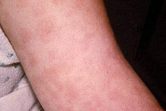
MACULE: A flat circumscribed area of discoloured skin (CDC)
PAPULE: Circumscribed skin elevation <0.5 cm (CDC/Dr Robinson)

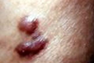
NODULE: Circumscribed lump >0.5 cm (CDC/Dr Steve Kraus)
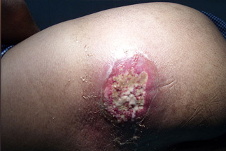
PLAQUE: Circumscribed disc-shaped elevation: <2 cm = small, >2 cm = large (CDC/Gavin Hart)

VESICLE: Visible collection of fluid <0.5 cm (CDC/Dr Noble)

BULLA: Visible collection of fluid >0.5 cm (CDC/Dr John Noble Jr)

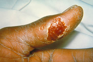
PUSTULE: Visible accumulation of pus (CDC/Brian Hill)

ULCER: Loss of epidermis (CDC/Dr Sellers)
WHEAL: Circumscribed area of cutaneous oedema (CDC/Dr William Foege)

There are many other terms used in dermatology. Some of these other terms will be useful in emergency medicine.
ABSCESS: Localised collection of pus (CDC/Bruno Coignard and Jeff Hageman)

ANNULAR: Ring-shaped (CDC/Renelle Woodall)

BURROW: Tunnel in the skin caused by a parasite e.g. scabies (CDC)

CARBUNCLE: Collection of furuncles (Wikimedia/Drvgaikwad)
CRUST: Dried exudate (CDC/Dr. Dancewiez)

CYST: Epithelial lined cavity filled with fluid or semi-solid matter (Wikimedia/Steven Fruitsmaak)

ERYTHEMA: Redness of the skin caused by vascular dilatation (CDC/M.A.Parsons)

EXCORIATION: Superficial abrasion due to scratching (Wikimedia/Berteun)

FOLLICULITIS: Inflammation of hair follicles (Wikimedia)

FURUNCLE: Pyogenic infection of the hair follicles (Wikimedia/Mahdouch)

LICHENIFICATION: Chronic thickening of the skin caused by rubbing or scratching (CDC)

PETECHIA: Haemorrhagic spot 1-2 mm diameter (CDC)

PURPURA: Extravasation of blood causing red discolouration of the skin or mucous membranes (CDC)

Telangiectasia: Dilated dermal blood vessels causing visible lesion (CDC/Robert E. Sumpter)

There are several serious conditions in dermatology that need to be immediately recognised. Fortunately for patients they are all relatively uncommon, however this may mean that you have never seen them before and might not immediately consider them as a diagnosis. This makes it even more vital that there is awareness in the ED of these medical emergencies.
Melanoma


Systemic features include:
- Arthritis
- Myalgia
- Palpitations
- Bell’s palsy
- Meningitis
- Polyneuropathy
- Psychosis
Image reproduced with permission from CDC/James Gethany.
- Graham-Brown R, Burns T. Lecture Notes: Dermatology. 8th edn. Blackwell Publishing, 2002: 9-12.
- Marks JG Jr, Miller JJ. Principles of Dermatology. 4th edn. Saunders Elsevier, 2006: 15-33.
- Gawkrodger DJ. Dermatology: An Illustrated Colour Text. 3rd edn. Churchill Livingstone, 2002: 14-19.
- Collier J, Longmore M, Duncan-Brown T. Oxford Handbook of Clinical Specialties. 5th edn. Oxford: Oxford University Press, 1999: 576-593.
- Gawkrodger DJ. Dermatology: An Illustrated Colour Text. 3rd edn. Churchill Livingstone, 2002: 45.
- Graham-Brown R, Bourke J, Cunliffe T. Dermatology Fundamentals of Practice. Mosby Elsevier, 2008: 208-209.
- National Institute for Health and Care Excellence (NICE). Meningitis (bacterial) and meningococcal septicaemia in under 16s: recognition, diagnosis and management. Clinical guideline [CG102], 2010. Last updated: 01 February 2015. [Accessed June 2023].
- Walls, Hockberger and Gausche-Hill. Rosen’s Emergency Medicine Concepts and Clinical Practice. 9th edn. Elsevier, 2017.
We should like to thank the following for permission to reproduce images:
- Centre for Disease Control
- Wellcome Images

Trek Tragedy!! Review on Lyme Disease
Lyme disease is a spirochete infection transmitted by ticks. ED presentation, although rare, is important to identify to enable early treatment.

Do not be rash with rashes: a guide to dermatological description
There are hundreds of skin diseases, many present with (often similar looking) rashes. Diagnosis of skin conditions can be a challenging task for non-dermatologists.

Common Childhood Exanthems
Children commonly present to the emergency department with a febrile illness and a rash
2 Comments
Very helpful
very useful and concise