Author: Richard Freeman / Editor: Lauren Fraser / Codes: NeoC5, SLO5 / Published: 18/03/2021
- Neonates can present with normal physiology to the paediatric emergency department. Studies have suggested that 1.9% of all patients present within the first month of life1.
- The most common diagnoses are the normal newborn (33.9%), indirect hyperbilirubinaemia (13.2%) and colic (5.8%)1. As such, a large proportion of these patients could be discharged without the need for paediatric review.
- During this session we will explore common benign conditions that could be managed by the emergency physician with relevance to the published evidence and national guidelines.

In this second part we will concentrate on three key areas:
- Expulsions from the upper GI tract
- Infantile colic
- Expulsions from the lower GI and GU tract
Basic science and pathophysiology
- Vomiting and feeding difficulties account for up to 36% of neonates presenting to the ED1.
- Visible regurgitation of feeds2:
- Is a common physiological event (affects at least 40% of infants)
- Usually begins before 8 weeks of age
- May be frequent (5% have six or more episodes per day)
- Does not usually require investigation or treatment
- Is also known as gastro-oesophageal reflux (GOR)
- Serious pathology is unlikely in well, hydrated infants without concerning features in the history or examination3.
Clinical assessment and risk stratification
GOR is likely when:
- There are no symptoms of serious illness (including sepsis).
- The child appears well with a normal examination.
- The vomiting is non-bilious and non-projectile.
- The child has plenty of wet and dirty nappies.
- The child is thriving.
- There are no features of gastro-oesophageal reflux disease (GORD):
- This is GOR that causes symptoms severe enough to merit medical treatment or reflux-associated complications2
A note on thriving:
- It is normal for a neonate to lose weight within the first few weeks of life especially if breast-fed.
- A general rule of thumb is no more than 10% of the birth weight and should be regained by day fourteen.
- However, this is based upon factors that predispose to jaundice and is highly controversial5.
- Recent evidence would suggest over 25% of otherwise healthy breast-fed neonates exceed these limits6.
- After this initial period insufficient growth should be determined by comparing the patient’s birth weight to the current weight on a growth curve.
Symptoms suggestive of serious illness:
- Bilious vomiting:
-
- 23-38% of neonates admitted with green vomitus were shown to have a surgical obstruction8,9
- It is important to ask about the colour of the vomit rather than use the term “bilious,” as most parents equated bile with the colour yellow7
- Projectile vomiting:
-
- 66-84% of cases will have pyloric stenosis3
- Poor urine and/or stool output:
-
- May indicate dehydration
- Excessive weight loss or pathological jaundice:
-
- May indicate dehydration

Signs suggestive of serious illness:
- Abnormal vital signs (including fever >38°C).
- Failure to thrive.
- Pathological jaundice (see previous module).
- Signs of raised intracranial pressure:
- Bulging fontanelle, rapidly increasing head circumference and sunset eyes
- Hence, head circumference should be considered part of the standard examination
- Signs of dehydration:
- Sunken fontanelle, poor capillary refill and decreased skin turgor

Signs suggestive of serious illness:
- Distended abdomen:
- 8% of full-term new-borns with abdominal distension have a congenital malformation (including congenital megacolon, anal atresia, malrotation, and intestinal atresia)10
- Hepatomegaly:
- May indicate inborn errors of metabolism
- An olive-sized mass in the right upper quadrant:
- Reported in 50% to 83% of cases of pyloric stenosis11,12
- Groin lump:
- May indicate an incarcerated inguinal hernia
Features suggestive of GORD:
- Marked distress:
- Currently defined as outside the normal range by an appropriately trained healthcare professional
- However:
- There is no persuasive evidence that prolonged crying or waking at night is related to GORD and there are other potential explanations2
- There is some evidence that abnormal posturing may be more suggestive especially if there are features of Sandifer’s syndrome2
- Episodic torticollis with neck extension and/or rotation that may be mistaken for seizure activity
- Apnoea:
- Observation studies suggest apnoea and GOR are rarely associated unless overt regurgitation is associated with the episodes2
- Hence, other causes of acute life threatening events should be excluded beforehand
- Feeding difficulties:
- Feed refusal, gagging and choking
- As with apnoea, observational studies suggest little evidence to support feeding difficulties are linked with GOR unless overt regurgitation is associated with the episodes2
- Faltering growth:
- Observational studies are highly variable with regards to the association between failure to thrive and GORD2
- However, overall consensus would suggest that faltering growth could be related to GORD however, other causes should be excluded first2
- Chronic cough/hoarseness of voice:
- Observational studies suggest no association between GOR and laryngeal inflammation in children2
- In the absence of associated overt regurgitation the presence of chronic cough or hoarse voice does not indicate the presence of GOR
- Complications:
- Reflux oesophagitis
- Upper GI bleeding, unexplained iron-deficiency anaemia, dysphagia
- Recurrent aspiration pneumonia
- Single episodes of pneumonia are relatively common in childhood however, consider if recurrent
- Reflux oesophagitis
- Frequent otitis media:
- Studies have demonstrated refluxate in the middle ear due to the presence of the digestive enzyme pepsin13,14
- Hence, frequent middle ear infections should raise the possibility of reflux2
Management
Investigations
- Serious pathology is unlikely in well, hydrated infants without concerning features in the history or examination3.
- Otherwise investigations should be used based upon the specific situation (i.e. septic screen, upper GI contrast study, USS abdomen).
Management: evidence base
- Positioning:
- There is evidence that prone and left lateral positioning is effective at reducing GOR in infants when measured by pH study2
- Useful when infants are awake and supervised
- However, when asleep the child should not be placed prone as the potential benefit is outweighed by the real risk of SIDS
- Feeding changes:
- One low quality comparative study found smaller volume feeds were associated with fewer reflux episodes when measured by pH monitoring15
- Thickened feeds:
- Fourteen comparative studies showed that thickened feeds reduced overt regurgitation and reflux acid exposure in infants2
- Alginates:
- Acts as a raft on the top of the stomach contents
- Three small RCTs comparing alginates to placebo suggested improvement in pH studies16-18
- Only one suggested an improvement in overt regurgitation19
- However, highly variable quality, Gaviscon formulation and different study ages prevent meta-analysis
- Proton pump inhibitors:
- Three RCTs reported no significant difference in reflux reduction when compared with placebo20-22
- However, two RCTs did find statistically significant reduction in reflux events21,23
- Studies very low to moderate quality
- H2 receptor antagonists:
- One RCT reported reduction in overt regurgitation when compared to placebo but not to statistical significance24
- Two RCTs reported outcomes relating to the resolution of oesophagitis or improvement in histology scores24,25
- Studies very low to low quality
- H2RA vs. PPI:
- Evidence from one very low quality RCT found no difference in outcome between PPIs and H2 receptor antagonists, but both improved symptom scores25
- Prokinetics:
- Increase gastric emptying
- One RCT found a statistically significant reduction in overt regurgitation26
- Two RCTs reported reduced acid reflux episodes based on 24 hour pH monitoring27,28
- Two RCTs found no difference in acid reflux episodes29-30
- Studies very low to moderate quality
- Prokinetics:
Adverse effects:
-
- Risk of extrapyramidal disorders and tardive dyskinesia with metoclopramide
- Small risk of ventricular arrhythmia and sudden cardiac death with domperidone
Management: general principles2
- Most require reassurance and safety-net advice only.
- The child should be placed on their back to sleep to reduce risk of SIDS.
- Parents should return if:
- Persistently projectile, haematemesis or bilious vomiting
- New concerns (marked distress, feeding difficulties or faltering growth)
- Persistent, frequent regurgitation beyond the first year of life
- Treatment nor investigation should not be offered for isolated overt regurgitation.
- Treatment nor investigation should not be offered for isolated;
- Unexplained feeding difficulties
- Distressed behaviour
- Faltering growth
- Chronic cough
- Hoarseness
- Single episode of pneumonia
- Hence, only treat if frequent regurgitation and marked distress.
Management: step-wise approach2
- Formula fed infants:
- Review the feeding history
- Reduce feed volume if excessive (see next section)
- Then, offer smaller more frequent feeds
- Finally, offer a trial of thickened formula
- Review the feeding history
- Breast fed infants:
- Breast feeding assessment by a person with appropriate expertise
Management: if step-wise approach fails2
- If formula fed, stop thickened formula.
- Offer an alginate (i.e. infant Gaviscon®) trial for 1-2 weeks.
- Continue if successful but try stopping at intervals to see if the infant has recovered.
- If cow’s milk allergy suspected:
- Elimination of cow’s milk from the diet for 2-3 weeks
- Maternal diary-free diet or nutramigen formula
- If symptoms resolve the diagnosis is highly likely
- NB: primary lactose intolerance (i.e. congenital absence of lactase enzyme) is extremely rare so do not offer lactose-free or other formula.
Management: if alginates fail2
- Consider a four week trial of a PPI or H2 receptor antagonists.
- Do not offer prokinetics without seeking specialist advice.
Management: when to refer acutely2
- Haematemesis
- Melaena
- Dysphagia
- Persistent, faltering growth associated with overt regurgitation
- Feeding aversion and a history of regurgitation
- Unexplained iron-deficiency anaemia
- A suspected diagnosis of Sandifer’s syndrome
Prognosis
- 90% of affected infants will be asymptomatic by one year of age2.
Basic science and pathophysiology
- A very common diagnosis that can also present with isolated regurgitation.
- Typically an infant requires 150ml/kg/day to ensure adequate growth.
- A fluid ounce is approximately 30ml.
- This is more difficult to assess in breast-fed infants but may be apparent with a careful feeding history.
- However, in both types of infant growth will be excessive.
Management
- If excessive feeding is suspected then this should be reduced to acceptable levels.
- Parents should be advised to return if:
- Persistently projectile, haematemesis or bilious vomiting
- New concerns (marked distress, feeding difficulties or faltering growth)
- Persistent, frequent regurgitation beyond the first year of life
Clinical assessment and risk stratification
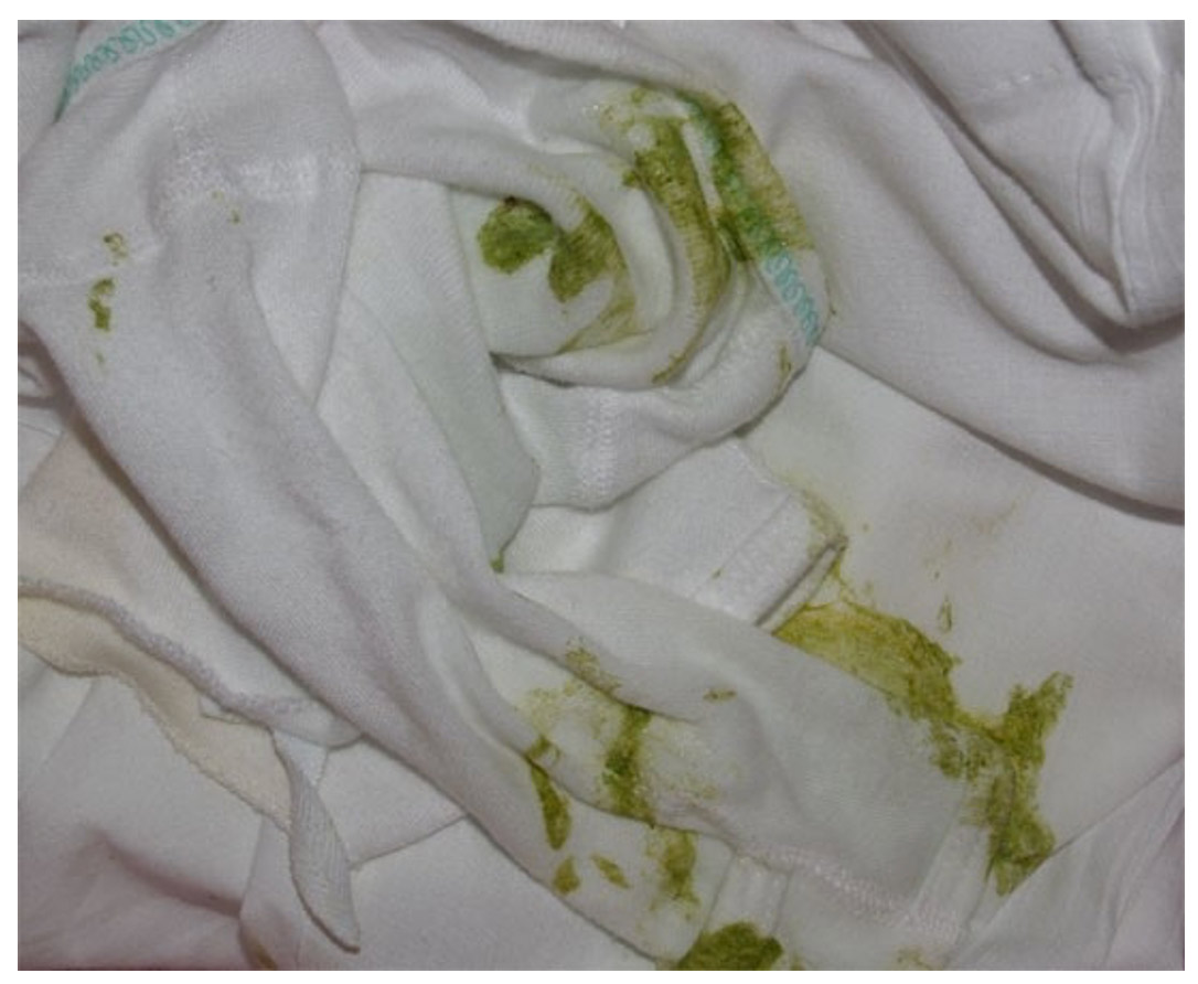
- Colostrum feeds within the first two days of life can produce bright yellow vomit shortly after feeding.
- The colour will be similar to that of the mother’s milk.
- Obviously this will not occur in bottle-fed infants and the child will remain well.

Management
- Reassurance is all that is required.
- Parents should be advised to return if:
- Persistently projectile, haematemesis or bilious vomiting
- New concerns (marked distress, feeding difficulties or faltering growth)
- Persistent, frequent regurgitation beyond the first year of life
- If there are any doubt the patient should be referred for a surgical opinion.
Clinical assessment and risk stratification
- Hematemesis in a healthy new-born is most often caused by swallowed maternal blood during delivery31-32.
- After the first 24 hours cracked nipples during breastfeeding is the most common cause31-32.
- Obviously this should not occur in bottle-fed infants after the first 24 hours.
- Differential diagnoses in “well” neonates include coagulopathy (especially if the infant did not receive a full dose of vitamin K) and congenital vascular lesions. In “sick” neonates consider DIC and liver failure.

- What is a full dose of vitamin K?
- A single intramuscular dose into the thigh given shortly after birth
- OR three oral doses given within the first month of life
- If any are omitted the infant is at risk of haemolytic disease of the newborn (HDN)
Management
- If an obvious source of maternal blood is found reassurance is all that is required.
- Parents should be advised to return if the child is unwell or they are concerned.
- If there are any doubt the patient should be referred for a paediatric opinion.
Basic science and pathophysiology
Epidemiology
- Colic is very common affecting 10-30% of infants33.
- Both sexes are affected equally.
- Feeding strategy does not appear to affect incidence.
- Preterm infants and those small for gestational age appear to be at increased risk34.

Aetiology
- Research has yet to identify the underlying cause.
- Suggested aetiologies include:
- Abnormal gastrointestinal motility and pain signals from sensitised pathways in the gut viscera35
- Inadequate amount of lactobacilli and increased concentration of coliforms within the gut flora36
- Behavioural issues such as family tension, parental anxiety or inadequate parent-infant interaction37

Image caption: Lactobacilli
Clinical assessment and risk stratification
Presentation
- Definition:
- Unexplained episodes of irritability, fussing or crying that last ≥3 hours per day on ≥3 days per week for ≥1 week38
- Hence, it is a diagnosis of exclusion.
- The typical symptoms are non-specific:
- Inconsolable crying (typically, high-pitched and occurring frequently in the afternoon or evening)
- Redness of the face
- Drawing up of the knees
- Flatus
- In-between episodes the infant remains well and thriving
Differential diagnosis
- In the acutely distressed infant consider:
- Physical discomfort (i.e. cold, wet, hungry)
- Medical:
- Sepsis
- Severe nappy rash
-
- Surgical:
- Intussusception (especially if facial pallor instead of redness)
- Volvulus (especially if billious vomiting)
- Strangulated hernia
- Testicular torsion
- Injuries:
- Corneal abrasion
- Non-accidental injury
- Hair tourniquets (digits, penis)
- Surgical:

Image caption: USS image of an intussusception showing the classical target
- If symptom are severe and recurrent consider:
- GORD (only if associated with regurgitation)
- Constipation or dyschezia
- Cow’s milk protein intolerance (CMPI)
- Parental distress will also exaggerate symptoms (consider maternal postnatal depression, inadequate support or ineffective parenting skills)
- NB: Remember isolated distressed behaviour is not a sufficient reason to investigate or treat for reflux as per the NICE guidelines2.
Management
Investigation
- The diagnosis is usually achieved from a typical history and a normal examination.
- Further investigations are not usually required.
Treatment
- Often reassurance is all that is required however:
- Living with a colicky baby can be exhausting for the family and hence, it is important to be empathetic and supportive
- Parents may be advised to share childcare until this stage has passed, in order to prevent physical/mental exhaustion
- If CMPI is suspected a two week trial of a diary free diet for breastfeeding mothers37 or nutramigen formula39 may be diagnostic
- If frequent regurgitation and marked distress consider treatment for GORD2
- There is no conclusive evidence for other interventions including soya milk, chiropractic care and massage
- There is little evidence to support the use of drugs such as Infacol®37 however they are unlikely to be harmful40
Prognosis
- Most infants with recover spontaneously by 3-4 months of age.
- However, it remains a frustrating problem for parents and it is important to support them through this challenging time.
- Cry-sis is a registered charity that offers support for families with excessively crying, sleepless and demanding babies. They also provide a telephone helpline between 9am to 10pm seven days a week.
Context
- Normal neonatal stools
- Neonatal constipation and dyschezia
- Urinary urate crystals
- Vaginal bleeding and leukorrhoea

Basic science and pathophysiology
Normal neonatal stools
- In general there is no such thing as a “normal” bowel habit in a neonate!
- It is completely normal to produce stool multiple times a day or only once a week especially if breast-fed.
- However, knowledge of how these stools change over the first week of life can provide useful diagnostic information:
- Meconium
- Transitional stool
- Yellow stool
a) Meconium
- Dark (almost black), sticky and odourless.
- Present in both breast and bottle-fed infants.
- Should first be passed within the first 48 hours of life.
- Otherwise consider Hirshsprung’s disease or meconium plug (may be associated with cystic fibrosis)
- Should convert to transitional stools by day four of life.
- Otherwise inadequate feeding should be considered

b) Transitional stools
- Should appear from day four of life if adequate feeding.
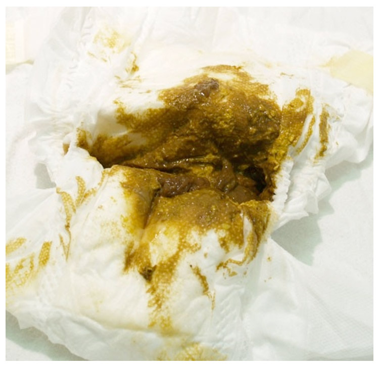
- Combination of dark coloured meconium with that of yellow stools characteristic of exclusive milk feeds.

c) Yellow stools
- Appear by day five in established breast-fed infants.
- Typically described as bright yellow and “seedy”.
- The typical consistency has significant liquid stool within the nappy with solid material on top and is incorrectly reported as diarrhoea by parents.
- In bottle fed babies the colour of established stool can vary between yellow, brown or green and has a thicker consistency similar to peanut butter.

Neonatal constipation and dyschezia
- It is completely normal for breast-fed infants to defaecate only once a week.
- Difficulty in passing stools is a common problem in neonates but rarely is it constipation.
- Healthy infants (<6 months) can strain and become distressed but subsequently pass soft This is a normal part of development as the child learns to coordinate increasing intra-abdominal pressure with relaxation of the pelvic floor. This is called dyschezia and will improve spontaneously overtime.
- Only if the stool is hard is it true constipation.
- True constipation in neonates is more suggestive of an underlying medical condition and these patients should be referred.
- The following should be considered:
- Hirschsprung’s disease
- Congenital anorectal malformations
- Spinal cord abnormalities
- Meconium ileus (cystic fibrosis)
- Metabolic causes: hypothyroidism, hypercalcaemia
Urinary urate crystals
- Inability to concentrate and acidify urine within the neonatal period facilitates a large urinary uric acid load41.
- The uric acid forms characteristic pink-orange deposits within the nappy of an otherwise well child.
- It is especially common within the first week of life.
- However, they can appear anytime within the first year as renal tubular function gradually improves.
- No investigation or treatment is required.

Vaginal bleeding and leukorrhoea
- High levels of maternal oestrogens within the new-born can lead to a physiological white-yellow discharge called leukorrhoea.
- As these levels fall (typically around day three) it is not uncommon for small withdrawal bleeds to occur.
- Both phenomenon resolve spontaneously by the first week of life as maternal hormones are metabolised.
- Large amounts of blood and bleeding after one week of age is atypical and alternative diagnoses such as coagulopathy and non-accidental injury should be considered.

Image caption: Oestrogen withdrawal bleed
Pitfalls
- Remember to work within your own competencies and follow local guidelines
- If the child appears unwell or you are unsure of the diagnosis you should always refer to your local friendly paediatrician or paediatric surgeon!
Session key points:
- In vomiting neonates serious pathology is unlikely in well, hydrated infants without concerning features in the history or examination (EL3a).
- Hematemesis in a healthy new-born is most often caused by swallowed maternal blood (EL4).
- GOR is very common and neither treatment nor investigation should be offered for isolated overt regurgitation (EL1a).
- For infants presenting with typical symptoms of colic and a normal examination further investigations are not usually required (EL4).
- There is little conclusive evidence for any medical intervention in patients with colic (EL1a).
- There is little conclusive evidence for use of drugs in the treatment of GORD (EL1a) but these should be introduced in a stepwise manner (EL5).
References
- Flanagan CF, Stewart M. Factors associated with early neo- natal attendance to a paediatric emergency department. Arch Dis Child 2014;99(3):239-243.
- National Institute for Health and Care Excellence. Gastro-oesophageal reflux disease in children and young people: diagnosis and management. NICE guideline [NG1] 2015.
- Ratnayake K, Kim TY. Evidence-based management of neonatal vomiting in the emergency department. Pediatr Emerg Med Pract 2014;11(11):1-20.
- Flaherman VJ et al. Early Weight Loss Nomograms for Exclusively Breastfed Newborns. Pediatrics 2015;135(1):e16-e23.
- Macdonald PD et al. Neonatal weight loss in breast and formula fed infants. Arch Dis Child Fetal Neonatal Ed 2003;88:F472-F476.
- Flaherman VJ et al. Early Weight Loss Nomograms for Exclusively Breastfed Newborns. Pediatrics 2014, peds.2014-1532.
- Walker GM et al. Colour of bile vomiting in intestinal obstruction in the newborn: questionnaire study. BMJ 2006;332(7554):1363.
- Malhotra A, Lakkundi A, Carse E. Bilious vomiting in the newborn: 6 years data from a Level III Centre. J Paediatr Child Health 2010;46(5):259-261.
- Godbole P, Stringer MD. Bilious vomiting in the newborn: how often is it pathologic? J Pediatr Surg 2002;37(6):909-911.
- Chen A, Du J, Du LZ. Clinical characteristics of abdominal distention in early newborns. Zhongguo Dang Dai Er Ke Za Zhi 2013;15(12):1074-1078.
- Shaoul R et al. Clinical presentation of pyloric stenosis: the change is in our hands. Isr Med Assoc J 2004;6(3):134-137.
- Benson CD, Lloyd JR. Infantile pyloric stenosis. A review of 1,120 cases. Am J Surg 1964;107:429-433.
- Aydin E et al. Role of nasopharyngeal reflux in the etiology of otitis media with effusion. Journal of Otolaryngology 2011;40:499-503.
- O’Reilly RC et al. The role of extraesophageal reflux in otitis media in infants and children. Laryngoscope 2008:118:1-9.
- Sutphen JL, Dillard VL. Effect of feeding volume on early postcibal gastroesophageal reflux in infants. Journal of Pediatric Gastroenterology and Nutrition 1998;7:185-188.
- Buts JP, Barudi C, Otte JB. Double-blind controlled study on the efficacy of sodium alginate (Gaviscon) in reducing gastroesophageal reflux assessed by 24 h continuous pH monitoring in infants and children. European Journal of Pediatrics 1987;146:156-158.
- Forbes D, Hodgson M, Hill R. The effects of gaviscon and metoclopramide in gastroesophageal reflux in children. Journal of Pediatric Gastroenterology and Nutrition 1986;5:556-55.
- Del, Buono R et al. Effect of Gaviscon Infant on gastro-oesophageal reflux in infants assessed by combined intraluminal impedance/pH, Archives of Disease in Childhood 2005;90:460-463.
- Miller S. Comparison of the efficacy and safety of a new aluminium-free paediatric alginate preparation and placebo in infants with recurrent gastro-oesophageal reflux. Current Medical Research and Opinion 1999;15:160-168.
- Hussain S et al. Safety and Efficacy of Delayed Release Rabeprazole in 1- to 11-Month-Old Infants With Symptomatic GERD. Journal of Pediatric Gastroenterology and Nutrition 2014;58:233-243.
- Omari TI et al. Effect of omeprazole on acid gastroesophageal reflux and gastric acidity in preterm infants with pathological acid reflux. Journal of Pediatric Gastroenterology and Nutrition 2007;44:41-44.
- Orenstein SR et al. Multicenter, double-blind, randomized, placebo-controlled trial assessing the efficacy and safety of proton pump inhibitor lansoprazole in infants with symptoms of gastroesophageal reflux disease. Journal of Pediatrics 229;154:514-520.
- Davidson G et al. Efficacy and Safety of Once-Daily Esomeprazole for the Treatment of Gastroesophageal Reflux Disease in Neonatal Patients. Journal of Pediatrics 2013;163:692-698.e2.
- Simeone D et al. Treatment of childhood peptic esophagitis: a double-blind placebo-controlled trial of nizatidine, Journal of Pediatric Gastroenterology and Nutrition 1997;25:51-55.
- Cucchiara S et al. Cimetidine treatment of reflux esophagitis in children: an Italian multicentric study, Journal of Pediatric Gastroenterology and Nutrition 1989;8:150-156.
- Leung AKC, Lai PCW. Use of metoclopramide for the treatment of gastroesophageal reflux in infants and children, CURR THER RES, CLIN EXP 1984;36:911-915.
- Bines JE et al. Efficacy of domperidone in infants and children with gastroesophageal reflux. Journal of Pediatric Gastroenterology and Nutrition 1992;14:400-405.
- Tolia V et al. Randomized, prospective double-blind trial of metoclopramide and placebo for gastroesophageal reflux in infants. Journal of Pediatrics 1989;115:141-145.
- Carroccio A et al. Domperidone plus magnesium hydroxide and aluminum hydroxide: a valid therapy in children with gastroesophageal reflux. A double-blind randomized study versus placebo. Scandinavian Journal of Gastroenterology 1994;29:300-304.
- Bellissant E et al. The triangular test to assess the efficacy of metoclopramide in gastroesophageal reflux. Clinical Pharmacology and Therapeutics 1997;61:377-384.
- Apt L, Downey WS. Melena neonatorium: the swallowed blood syndrome. J Pediatr 1955;47:6–12.
- McCollough M, Sharieff GQ. Common complaints in the first 30 days of life. Emerg Med Clin North Am 2002;20:27–48.
- Savino F et al. Looking for new treatments of Infantile Colic. Ital J Pediatr 2014;40:53.
- Milidou I et al. Gestational age, small for gestational age, and infantile colic. Paediatr Perinat Epidemiol 2014;28(2):138-45.
- Savino F, Tarasco V. New treatments for infant colic. Curr Opin Pediatr 2010;22(6):791-7.
- Chau K et al. Probiotics for infantile colic: a randomized, double-blind, placebo-controlled trial investigating Lactobacillus reuteri DSM 17938. J Pediatr 2015;166(1):74-8.
- Hall B, Chesters J, Robinson A. Infantile colic: a systematic review of medical and conventional therapies. J Paediatr Child Health 2012;48(2):128-37.
- Hyman PE et al. Childhood functional gastrointestinal disorders: neonate/toddler. Gastroenterology 2006;130(5):1519-26.
- Iacovou M et al. Dietary management of infantile colic: a systematic review. Matern Child Health J 2012;16(6):1319-31.
- Management of infantile colic. BMJ 2013;10;347:f4102.
- Passwell JH et al. Functional excretion of uric acid in infancy and childhood. Arch Dis Child 1974;49:878-82.
Further Reading
- Newborn Photo Gallery. Stanford School of Medicine [Online]. [Accessed 18th February 2021].
- National Institute for Health and Care Excellence. Gastro-oesophageal reflux disease in children and young people: diagnosis and management [NG1]. London: National Institute for Health and Care Excellence 2010.
- Cry-sis [Online]. [Accessed 18th February 2021].


3 Comments
Good
i agree alex bhai ! 🙂
Thank you. Some new and relative information to what we deal with this.